 Except the papers and where otherwise noted, this work is licensed under a Creative Commons Attribution 4.0 International License
Except the papers and where otherwise noted, this work is licensed under a Creative Commons Attribution 4.0 International License

Access the manuscript or click on the links below to visit the large-scale EM data at full resolution.
 |
 |
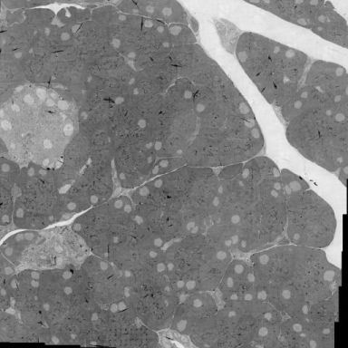 |
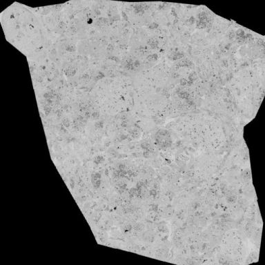
Fig. 2a. Large scale EM (human pancreas) using our workhorse single-beam STEM - Routine ‘nanotomy’as performed in our imaging facility. A cross-section of a human Islet imaged at 2.5 nm pixelsize with a single beam microscope (see de Boer et al. NatComm 2020 for details). |
Fig. 2b. Multi-beam nanotomy using a FAST-EM prototype* - Imaged at 4nm pixel size in 15 minutes. Images are produced by scanning all 64 beams simultaneously over a field of view, after which data is seamlessly stitched together to form a single image. Image courtesy of Delmic B.V., Delft, The Netherlands.* delmic.com/en/products/fast-imaging/fast-em |
Disclaimer - Contact - Home